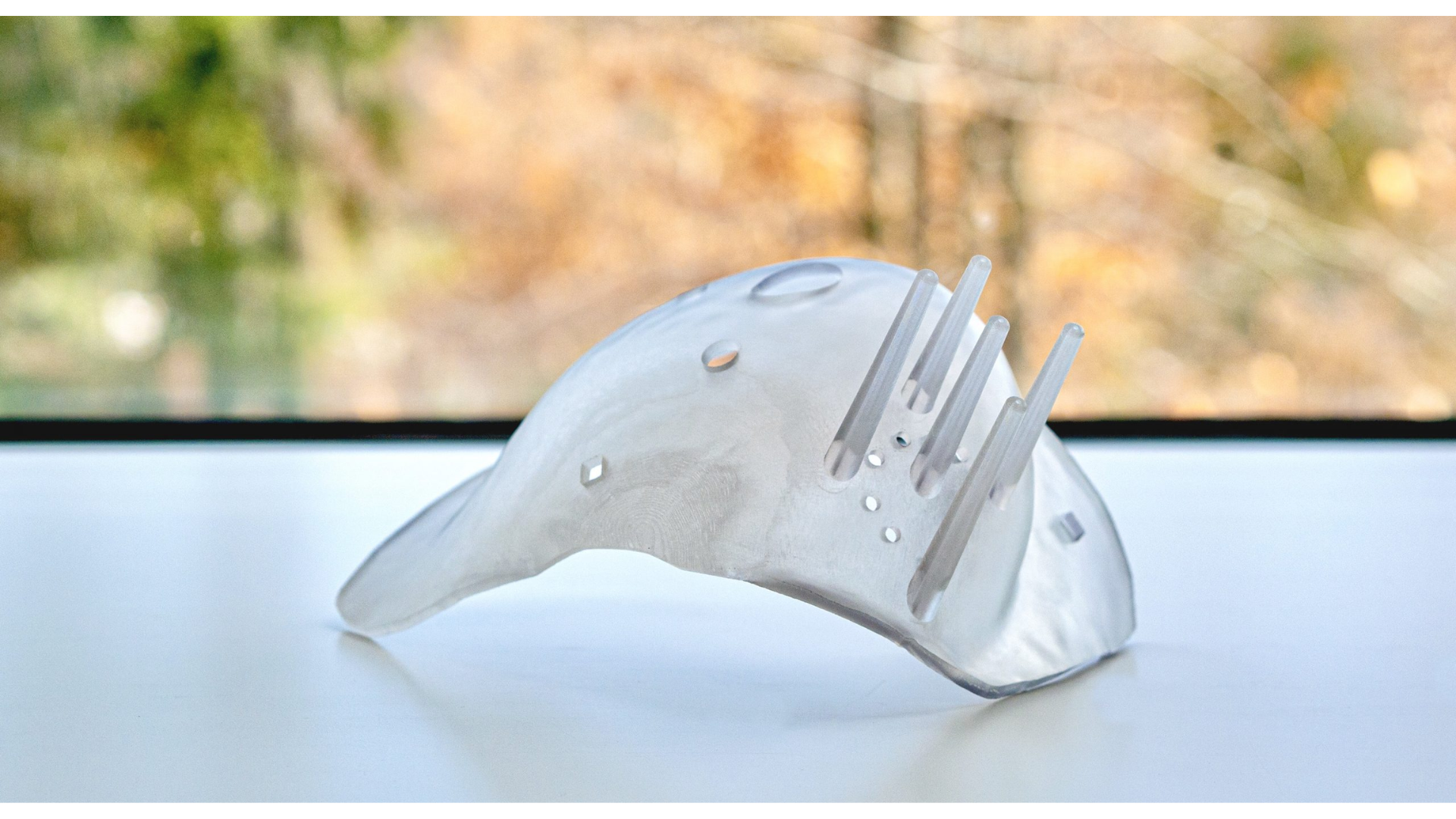
LEBANON, N.H./January 24, 2023 — CairnSurgical, Inc., an innovator striving to make breast cancer surgery more precise, announced that the first patients have been treated in its European post market clinical study of the 3D-printed Breast Cancer Locator (BCLTM) System. The successful first cases have been performed at Spital Zollikerberg in Zurich, Switzerland and Santa Chiara Hospital in Pisa, Italy. The BCL System is designed to improve the accuracy of breast conserving surgery (lumpectomy) by providing precise guidance regarding tumor shape, size, and location, thus enabling the ability to achieve clear margins.
“We are delighted that in our first case using the BCL technology we were able to achieve a negative margin, with no detectable cancer present after the breast tumor was removed. This is the result we strive to achieve for every patient,” said Prof. Dr. med. Hisham Fansa, head of the Breast Center and head of plastic surgery for Spital Zollikerberg, and co-principal investigator of the study. “We are excited about having more detailed 3D information about each tumor and a surgical guide that is personalized for each patient, which may help us make breast cancer surgery more accurate and thereby reduce the need for additional surgery for our patients.”
The multicenter European post market surveillance clinical study is being conducted in selected European countries within the MDR custom device guidelines. The study will take place in several centers in Europe, including Santa Chiara Hospital, Spital Zollikerberg and Agaplesion Markus Krankenhaus in Frankfurt, Germany. The primary endpoint is positive margin rate, which is assessed by imaging and pathology immediately following the procedure.
Current Breast Conserving Surgery (BCS) is unsuccessful at removing the entire tumor about 20 percent of the time, primarily because current tumor localization techniques do not provide the information required to achieve precise removal of the entire disease. According to a recent study on breast cancer tumor shapes, less than 20 percent are spherical in shape,1 making techniques that place implants or wires in one or two central points within the tumor of limited to no value in identifying its size, boundaries and margins.
The BCL System is designed to reduce positive margin rates, and to improve the process of BCS, using three steps.
- A supine MRI image is first performed, with the breast positioned in its surgical position, providing detail on the size, shape, and location of the tumor. This imaging data is used to analyze the tumor and construct a three-dimensional model that serves as the basis for two surgical tools.
- A 3D-printed form – the Breast Cancer Locator, or BCLTM – is produced to fit the unique shape of the patient’s breast, with ports for several central and bracketing wires to be placed under anesthesia in the operating room immediately prior to surgery. The wires guide the surgeon to the tumor boundaries and margins.
- An interactive 3D image of the tumor in the breast – the VisualizerTM – is provided to the surgeon, which can be manipulated during surgery. The Visualizer offers 360° views of the tumor and quantifies the closest distance from the tumor to the skin and chest wall – information that provides valuable references for the surgeon during the procedure.
“By personalizing a guidance solution for each individual patient’s tumor and breast, in 3D and using supine MRI data, we are enabling precision medicine for breast cancer patients,” said Richard Barth, Jr., MD, Dartmouth-Hitchcock Medical Center, and Professor of Surgery, Geisel School of Medicine at Dartmouth, and Co-founder, CairnSurgical. “With two multicenter clinical studies underway in the U.S. and Europe, we move ever closer to being able to make the BCL System accessible to surgeons who value precision and want to reduce repeat surgeries for their patients.”
The U.S. pivotal trial of the Breast Cancer Locator (BCL) System is currently enrolling patients and is designed to support FDA clearance and U.S. commercialization. The multicenter, 1:1 randomized, controlled trial encompasses 448 women treated at up to 25 centers. The primary endpoint is positive margin rate, which can be assessed immediately following the procedure. Other endpoints include specimen volumes, rate of additional shave biopsies, re-excision rate, cancer localization rate, operative time, and cost of care.
In two studies of the BCL System, published in the Annals of Surgical Oncology2 and in Surgical Oncology3, clear tumor margins were achieved in all 33 patients. Resection volumes were comparable to optimal resection volumes determined by tumor modeling based on the supine MRI.2,3
The Breast Cancer Locator is considered an investigational device in the U.S. and is limited by U.S. law to investigational use only.
1. Byrd BK, Krishnaswamy V, Gui J, et al. The shape of breast cancer. Breast Cancer Research and Treatment. 2020; 183: 403-410.
2. Barth RJ, Krishnaswamy V, Paulsen KD, et al. A patient-specific 3D-printed form accurately transfers supine MRI-derived tumor localization information to guide breast-conserving surgery. Ann Surg Oncol. 2017; 24(10): 2950-2956.
3. Barth RJ, Krishnaswamy V, Rooney T, et al. A pilot multi-institutional study to evaluate the accuracy of a supine MRI based guidance system, the Breast Cancer Locator™, in patients with palpable breast cancer. Surgical Oncology, Vol 44, September 2022, 101843.


